Opening hours Mo - Fr: 9.00 - 21.00 Saturdays: 9:00 - 15:00 | Phone22 620 21 40
  • pl
  • en
  • DENTAL
    SURGERY

    DENTAL SURGERYPiezosurgeryTooth extractionSurgical removal of impacted toothRoot canal treatmentApiocetomy (root end resection)Tooth hemisectionBone regeneration with biomaterialsSINUS LIFTRepair of oroantral communicationsSplittingLingual and labial frenulectomy / frenulotomyAlveoloplastyOncology diagnosticsFAQ

    Car racing is like tooth extraction. You need to know how to do it!
    Lewis Hamilton

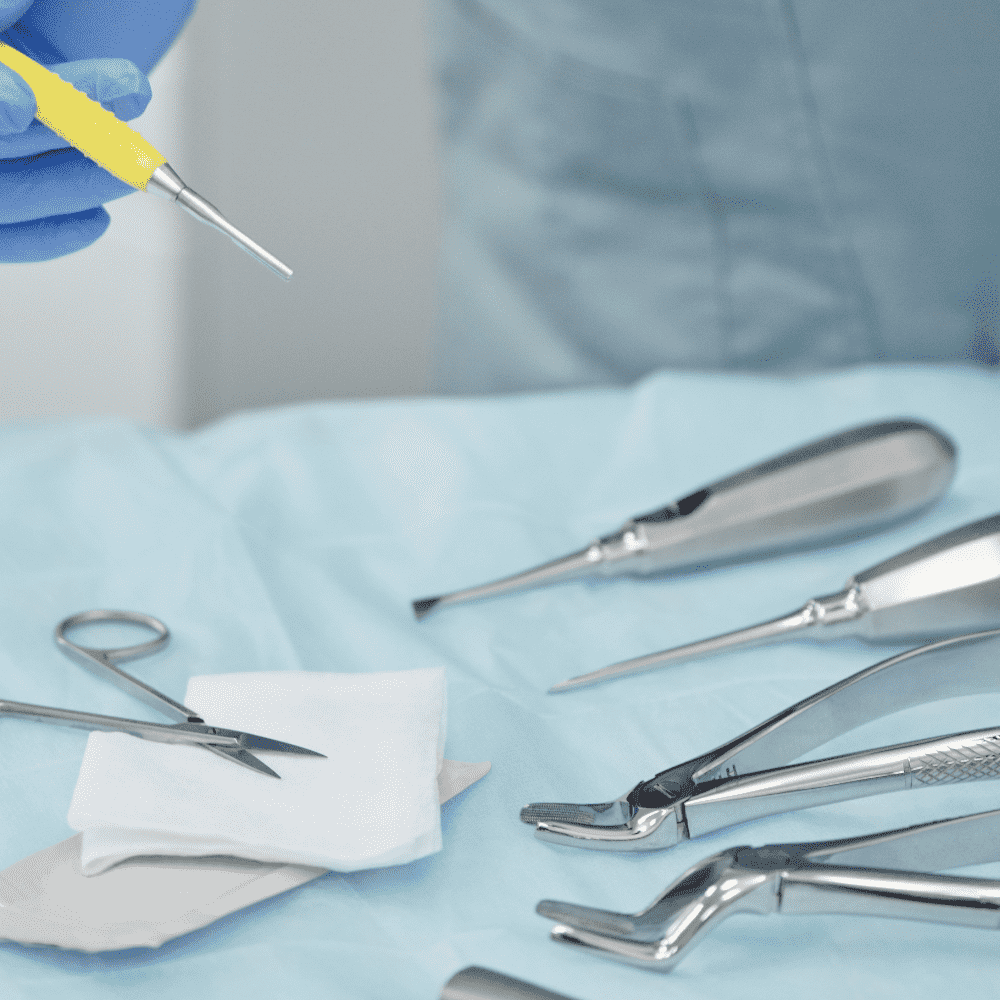
    Dental surgery is a field of medicine that deals with surgical treatment of oral cavity and adjacent areas. It emerged in the second half of the 19th century as a field of dentistry requiring general surgical training. As part of this discipline, a number of procedures involving the oral cavity are performed, including procedures supporting other areas of dentistry, such as endodontics – rood-end resection, periodontology–periodontal surgery, prosthetics – pre-prosthetic surgery, or the latest discipline – implantologyWe are happy to inform you that at Villa Nova Dental Clinic we are able to perform all surgical procedures inside the oral cavity and in adjacent areas, which do not require hospital treatment.

    We associate surgical procedures with pain and long periods of recovery. Nothing could be further from the truth. The rules of surgical treatment have changed significantly over the recent years. Modern surgery involves ATRAUMATIC teeth extraction that allows to retain as much healthy bone as possible, lost tissue restoration, as well as seamless surgical procedures.

    Thanks to state-of-the-art diagnostic devices and advanced materials at Villa Nova Dental Clinic, we are able to precisely plan and perform each dental surgical procedure.With the use of equipment such as Piezosurgery (an ultrasound device for cutting and splitting bone) and XO Odontosurge (a device using high frequency electric current used for bloodless treatment of soft tissue in the oral cavity), the procedures are minimally invasive. Painlessness is guaranteed byan advanced, computer-controlled injection device for giving local anaesthetic – The Wand (see-> our clinic is pain-free). We also offer the option of performing procedures under general anaesthesia.

    We need to do it with surgical precision.
    JF Kennedy

    Piezosurgery is a modern bone surgery technique used in procedures in the area of periodontics, implantology, as well as oral and maxillofacial surgery. The technique has significant advantages over traditional surgical techniques.

    Due to high level of accuracy (micrometric cuts) and the possibility of saving tissue (selective cuts), piezosurgery is characterised by exceptional precision and safety.

    Piezosurgical technique uses ultrasonic waves (subtle vibrations), which allows for cutting hard tissues while preserving delicate soft tissues. Thanks to this technique, the surgical site is virtually blood-free. Frequency regulation and a wide selection of surgical tips makes it possible to adjust the device to work with different surgical techniques, e.g. during:

    • tooth extraction,
    • sinus lift,
    • bone tissue procurement procedures,
    • splitting of alveolar process during implantation,
    • osteodistraction,
    • surgical procedures in the treatment of dental pulp diseases,
    • soft tissue cyst removal.

    Piezosurgery meets the requirements related to fitting dentures on dental implants. The precision of this procedure technique allows for perfect results, while tissue saving accelerates the healing process. For the patient, this means:

    • decreased oedema,
    • smaller post-surgical pain,
    • better comfort during the procedure itself.

    Tooth extraction is a surgical procedure consisting in removing a tooth from a dental socket with the use of appropriate instruments (forceps, levers etc.). The procedure is performed under anaesthesia, with aseptic surgical site (see -> our clinic is pain-free). Each tooth extraction procedure should be performed in an atraumatic way, i.e. in a way that the it causes minimal damage to the surrounding tissues, both hard (bone) and soft (gum). The dentist’s skills and co-operation of the patient ensure the appropriate course of the procedures and prevent complications.

    The most important indications for tooth extraction include chronic inflammation of the periapical tissue,severe tooth fracture, as well as advanced periodontitis.

    Please remember that tooth extraction is a LAST RESORT! At Villa Nova Dental Clinic we make every effort to avoid it, making use of the most advanced achievements in dentistry.

    RECOMMENDATIONS AFTER DENTAL EXTRACTION

    Directly after tooth extraction, it is absolutely necessary to maintain oral hygiene. In order to stop bleeding you should bite on a piece of sterile gauze for a certain period of time, usually 30 to 45 minutes. You are also not allowed to smoke for 24 hours after extraction. During this time you should avoid intensive mouth washing, as well as brushing teeth around the socket. You also shouldn’t drink any hot liquids.

    An impacted tooth is a tooth that remains unerupted despite being completely developed (it may be completely invisible in the oral cavity or partly visible). The reason for tooth impaction is the fact that it is being blocked by other teeth or that it grows in a wrong direction. This problem often concerns wisdom teeth. Surgical tooth extraction is a more complex procedure and requires more time than “regular extraction”.

    Thanks to our specialists’ skills, as well as the application of modern devices, such as Piezosurgery, surgical extraction of an impacted tooth is not a major nuisance for the Patient and is characterised by faster healing process.

    Indications for impacted teeth extraction include: planned orthodontic or prosthetic treatment, developing intrabony odontogenic cysts of significant scope, pressure on neighbouring teeth that causes the risk of root resorption, as well as inflammations and pain complaints.

    It should be borne in mind that impacted tooth extraction is a difficult procedure, carrying the risk of more potential complications due to the atypical location of the tooth, as well as unexpected difficulties that may arise during the procedure.

    Root canal treatment

    Root canal treatment (endodontics) is applied in cases of caries complicated by inflammation, necrosis or gangrene (disintegration) of the dental pulp.
    It is unfortunately very often the last chance of saving a tooth. Endodontic treatment consists the removal of infected or dead pulp from the pulp chamber and root canals, mechanical dilatation and disinfection of canals and tight filling of these spaces with special materials.
    It sounds quite simple, but without the endodontist’s specialist knowledge and experience, the necessary equipment and a number of other factors, it becomes “mission impossible”.
    Our endodontists deal with Patients who are referred from other clinics in order to try to save that teeth, which without the use of modern technologies, would be needed to extract.

    At our clinic, you will find only the best and the most modern dental equipment:
    – modern tools for canal preparation, both manual and mechanical (K3 ENDO, PROFILE, RC PROC, ultrasound tools);
    – Kofferdam – used to isolate saliva;
    – Endometers – RAYPEX 5, ENDOMASTER.
    – systems for filling the dental cavity (OBTURA II, SYSTEM B);
    – loupes and endodontic microscopes: CARL ZEISS and LEICA microscopes – thanks to these devices many teeth that would otherwise be removed can now be saved. By providing an enlarged image at great big depth, a wide field of vision and very good illumination of the examined area, the microscope allows the observation of details invisible to the naked eye. The application of the microscope is of particular assistance in seeking root canal openings and in locating and removing broken canal tools. A video camera included with the set allows image transmission and recording of the procedure. The correctness of the procedure performed may be immediately verified by an X-ray taken in the room (on the dentist’s chair) or in our X-ray laboratory.

    Root end resection is a procedure whereby a tooth’s root tip is removed, inflammatory lesions are treated and the root is protected against recurrent inflammation. This method of treatment enables normal bone restoration in place of the removed lesions and this process can be supported by using material that stimulates bone rebuilding. In most cases this procedure allows to save the tooth from extraction (95% effective). Due to an enormous progress in orthodontic treatment (endodontic microscopes, advanced devices for root preparation), such procedures are becoming rare.

    When apical lesions cannot undergo endodontic treatment or when repeated canal treatment is not an option, root end resection becomes the only available therapy.

    Root end resection procedure is indicated when:

    • it is impossible to carry out endodontic treatment that reaches up to the very end of the canal (abnormal anatomical structure, “overgrown” root canal);
    • the material filling the canal is extruded beyond the tip of the root, causing severe pain,
    • aesthetic and functional prosthetic restoration in the form of a crown-root inlay and a prosthetic crown has been made, and an inflammatory lesion developed around the tip of the root that cannot be treated in any other way;
    • the applied treatment fails to remove inflammatory lesions around the dental root.

    Root end resection is usually carried out on single-root teeth (incisors, canine teeth); however in practice, the procedure may be carried out on any tooth that requires such treatment

    Hemisection involvesseparation and removal of a part of the crown and root of a tooth affected by inflammation with irreversible pathological lesions.

    During the hemisection procedure the dentist removes the damaged crown of the multi-root tooth together with the corresponding crown. Hemisection is carried out on multi-rooted teeth (usually molars).

    With this procedure the dentist can keep a fragment of the tooth that can later serve as an abutment tooth in subsequent prosthetic reconstruction (see -> crowns).Hemisection also allows the dentist to prevent potential inflammatory lesion around the tip of one of the roots.

    Hemisection is a procedure carried out when conservative treatment is no longer possible. Indications for hemisection include:

    • apical lesions next to the roots
    • failure of endodontic treatment
    • vertical fracture of tooth and/or root

    CONSTANT DEVELOPMENT OF MODERN IMPLANTOLOGY ENABLES IMPLANT PLACEMENT EVEN IF THE PATIENT’S NATURAL BONE IS NOT SUFFICIENT IN A GIVEN SPOT.

    It is estimated that in around 40% of cases of implant treatment bone restoration and regeneration is required. It is possible thanks to the use of bone substitute biomaterials, enabling the restoration of lost bone structures.

    Guided bone regeneration allows for proliferation of bone tissue in the required location in order to appropriately place the implant with regard to future prosthetic restoration. The decision about the application of guided bone regeneration is made after a radiological analysis. Sometimes such necessity will only be revealed during the procedure, because routine X-ray will not always provide a full clinical image of the surgical site.

    Bone loss occurs:

    • after tooth extraction- with time the process of gradual loss of the denticle, both in the maxilla and the mandible (the optimal period for placing an implant is 3-6 months after tooth extraction);
    • after tooth loss due to periodontitis or mechanical injury;
    • after complex and traumatic extractions, when there is significant damage to the tissues surrounding the tooth;
    • in case of long-term use of partial and full dentures (due to the pressure of the denture on the bonethrough the gum, causing its degeneration).

    All cases described above require bone regeneration that helps achieve a satisfying functional and aesthetic effect.

    Guided bone regeneration is usually carried out together with implantation. Sometimes, in case of particularly large bone void and a lack of potential stability, bone regeneration is performed as a separate procedure, and implants are placed only after the bone tissue is formed – usually after 6-8 months.

    The procedure itself consist in fixing biomaterial in the space of the planned bone regeneration and shaping it in an appropriate way. Then, everything is covered with a collagen barrier membrane and stitched up. The application of a resorbable collagen barrier membrane prevents epithelial cell growth on biomaterials and enables bone tissue growth.

    At Elektoralna Dental Clinic we only use effective biomaterials (Bio-Oss, Endobon) and membranes (Bio-Gide, Osseo-Guard), which have been studied in clinical trials. They guarantee a very high proportion of successful procedures with long-lasting results.

    Implant treatmentis the best solution for replacing missing teeth. Unfortunately, in order to have an implant fitted, the patient’s alveolar process needs to be of an appropriate width and height. One of the limitations for the implantation procedure in the lateral maxilla is that the sinus is often lowered.

    This is due to the fact that with every year following the extraction of lateral teeth, the lateral bone height gradually decreases. The process is very slow, but continuous. When vertical bone height falls below 8~10 mm the sinus lift procedure becomes necessary. The procedure allows for the fitting of implants through reducing sinus volume,while increasing bone base necessary for the placement of dental implants.

    There are two sinus lift techniques:

    • open method– applied in a situation where the thickness of the existing  bone is less than 6-7 mm. The procedure starts with drilling a window in the lateral sinus wall. For opening the sinus, a piezoelectric bone cutting instrument is used, which ensures great precision of the procedure, as well as its atraumatic character (see -> Piezosurgery). After the mucous membrane is lifted, a bone substitute material is placed in the space obtained at the bottom of the sinus. The bone window is closed with a barrier membrane, covered with the patient’s mucous membrane (gum) and stitched tightly (see -> bone regeneration with biomaterials). Depending on the indications, the implant fitting procedure is carried out during the same appointment or after a period necessary for the formation of a new bone tissue.
    • closed method – applied where the thickness of the existing  bone exceeds 6-7 mm. The placing of the substitute bone material is carried out through a whole drilled for the purpose of fitting an implant. The sinus mucous membrane is elevated using special instruments – osteotomes – in order to create free space between the mucous membrane and the alveolar bone. Then the biomaterial is placed in this space and the implant is fitted during the same appointment. The disadvantage of this method is a limited scope of bone reconstruction, the necessity to repeat the same procedure with every implant that is fitted, lack of visual control over the surgical site, which significantly increases the risk of sinus mucous membrane perforation.

    THE PURPOSE OF SINUS LIFT IS TO RESTORE THE CHEWING FUNCTION AND TO IMPROVE THE PATIENT’S APPEARANCE, regardless of the causes and extent of alveolar degeneration in the lateral maxilla.

    Sometimes extraction of upper posterior teeth creates communication between the sinus and the oral cavity. This is because the root tips of these teeth are stuck in the lumen of the sinus. Ifan oroantral communication is diagnosed, a panoramic X-ray and/or  X-ray of paranasal sinuses is taken. Thenthe presence of a sinus inflammation, cyst or other material needs to be ruled out. If the conditions mentioned above are not diagnosed, the closure of oroantral communication is performed through removing gums and moving them in such a way that the passage to the sinus created after the tooth extraction is closed. The wound is sutured for around two weeks. During this period the patient needs to strictly follow the doctor’s recommendations and undergo radiographic sinus assessment for several weeks after the procedure.

    After a loss of tooth or dental extraction, the dental process begins to disappear. The narrow alveolar process in a limitation for the fitting a dental implant. Often in in such cases a prior bone regeneration of the lost tissue is required.

    In case of inadequate width of the alveolar process, but with adequate height, in order to place a dental implant correctly, the bone splitting technique needs to be applied.

    Split lamina create space that allows for stable placement of the dental implant between them. A supplement to this method is the application of a bone substitute preparation in the fissure. What proves very helpful in this method is piezosurgery, which guarantees an atraumatic procedure.

    Lip frenulum and tongue frenulum are folds of tissue that connect the lip or the tongue with the alveolar process. There are common abnormalities associated with the frenula, such asfrenulum located too close to the teeth, frenulum too short or thick,abnormalities that negatively affect the periodontium, the aesthetics of the teeth, the formation of a diastema, the stability of removable dentures, speech impediments or feeding disorders. These abnormalities can be corrected through procedures performed on the frenula: frenulectomy and frenulotomy.

    Frenulectomyis a simple incision of a too tight frenulum that is too tight, causing restrictions in movement of a tongue or lips.Frenulotomyconsists in removing a too thick or abnormally located frenulum, usually of the upper lip, and sewing it in the proper place. In case of a hypertrophic lingual frenulum very often functionality is impaired, leading to speech disorders or difficulties in taking in food.

    The procedures are carried out under local anaesthesia, with the use of special scissors or an electric knife, which eliminate the necessity of putting on sutures.

    Alveoloplasty is a surgical procedureinvolving the regeneration of the alveolar bone after dental extraction or tooth loss with the use of bone substitute material. Its purpose is to restore the original bone shape. It is usually performed before the planned prosthetic treatment with removable restorations in order to smooth out bone irregularities. Without it, the denture would be impossible to adjust and uncomfortable to wear for the Patient.

    Standard head and neck examinations should be performed by a dentist for each patient, at least once a year. It is always examination by signt and touch. These are not sufficient methods to detect cancer at an early stage. Oral cancer ranks 5th place among gastrointestinal neoplasms and 8th among all neoplasms. 70% of malignant tumors are diagnosed too late, in III or IV stage. To detect pathologies as ealry as possible, we use the Vizi Lite Plus test, to help indentify lesions when they are not visiable or tangible. If there are already visiable outbreaks it allows for their initial qualification and mornitoring.
    The examination is completely non-invasive.

    1. How should I prepare myself for the surgical procedure?

    To be prepared for the surgical procedure, you should:

    • take a rest;
    • have a light, but calorific meal;
    • take your regular medicines unless the dentist recommends otherwise;
    • relax and trust your dentist!

    2. What is allowed and what is forbidden after tooth extraction?

    After the tooth extraction procedure:

    • remove the dressing approximately 40 minutes after the procedure;
    • do not eat and drink anything for 2 hours after the procedure;
    • eat only light, semi-liquid food for the whole day after the procedure;
    • do not rinse your mouth;
    • put a cold compress on the face for 2-3 hours after the procedure (recommended);
    • do not warm your face on the side of the extracted tooth;
    • avoiding smoking on the extraction day would be of benefit;
    • in the case of pain you may use any OTC painkiller available, except products containing acetylsalicylic acid, e.g. Aspirin(R).

    3. Is it necessary to remove all the wisdom teeth?

    If the wisdom teeth are healthy, regularly positioned in the dental arch, do not pose difficulties in cleaning and do not cause pain, there is no need to remove them unless for other reasons, e.g. orthodontic indications. The following are indications for the extraction of wisdom teeth (including impacted ones):

    • uselessness in biting;
    • lack of space in the dental arch;
    • certain diseases of the dental pulp, such as purulent inflammation or gangrene;
    • bone pouch outside the tooth crown;
    • pressure on the neighbouring tooth;
    • cyst around the crown of the impacted tooth (visible on the X-ray);
    • neuralgia caused by the pressure of the tooth on a nerve.

    4. Is it advisable for teeth to be removed in pregnant women?

    The pregnancy itself is not a contra-indication for tooth extraction. However, the procedure should be appropriately scheduled, the patient should be appropriately prepared and the tooth should be removed quickly and efficiently. The best period for such procedures is the second trimester of the pregnancy. In the first trimester most of the procedures in the oral cavity to be performed are postponed, unless such delay would be a threat for the patient’s health or life. The most important element of the preparation of the patient for such a procedure is calming her and explaining the need for her tooth extraction. Only the most safe and efficient agent may be used for the anaesthesia of pregnant women.

    5. How long may a wound bleed after a surgical procedure?

    Each surgical procedure is associated with bleeding. The regular bleeding duration varies from several to some dozen minutes (until the clot is formed). An apparently longer bleeding period is considered abnormal, and there may be various underlying general (bleeding tendency, effect of medicines) or local (extra wound, bone injury) causes for such abnormality. Irrespective of the cause of such bleeding, the patient should visit the surgery immediately to seek help.

    6. What is the dry socket?

    The dry socket is a complication following tooth extraction consisting of the lack of a blood clot in the alveolus (a clot has not been formed or has been washed out), commonly associated with bacterial infection. The symptoms, involving severe sharp pain, radiating to the ears or temples and commonly occurring during the night, as well as general weakness, usually occur 2-3 days after the procedure. Such ailments may persist up to two weeks. The exact cause for such disease is unknown; possible causes include compromised immunity, vitamin deficiencies, smoking, etc. Should similar symptoms occur, the patient should seek help from a dental surgery. The treatment applied will accelerate the wound healing and relieve the pain.

    7. Can a knocked-out tooth be saved?

    If a tooth has been knocked out, it may be reimplanted, i.e. placed again in its alveolus. The success of such procedure depends of three factors:

    • the stage of development of the tooth; the best chance of successful reimplantation is with young teeth, whose roots have not developed completely;
    • the duration when the tooth was outside the alveolus (preferably not longer than 20-30 minutes);
    • The conditions in which the tooth was kept (it is recommended to keep the tooth in the patient’s mouth or in a container with milk or water.

    8. Is the sinus lift procedure painful?

    Modern dentistry doesn’t leave room for pain. In our surgery we offer several types of anaesthesia. This allows us to offer full and thorough anaesthesia and therefore all procedures are painless.

    9. Are implants placed at the same appointment as the sinus lift procedure?

    The decision about implants being fitted immediately or after a certain period is made by the doctor during the procedure planning and depends on several factors, such as bone thickness in the spot of planned implantation.

    10. After what time can the treatment be completed after the sinus lift procedure?

    Time required for implants to heal-in is the same as with standard implantation in the maxilla, i.e. about 6 months. After this period final crowns can be made and the treatment can be completed. In exceptional cases the period might have to be longer. This depends on patient’s anatomical conditions and the scope of the sinus lift procedure.

    11. How often is bone regeneration required?

    Statistically 40% of implantological procedures require bone regeneration. Failure to undergo bone regeneration despite indications may cause a deterioration of implant settlement within the bone and a decrease in the implant-bone contact surface.

    12. What is dry socket?

    Dry socket is a complication of dental extraction, which occurs when a blood clot fails to develop in the tooth socket (the clot failed to develop or has been washed out). It is often accompaniedby a bacterial infection. The symptoms involving severe sharp pain radiating to the ears or temples and commonly occurring during the night, develop 2-3 days after the procedure. Such ailments maypersist for around 2 weeks. The exact cause of the condition is unknown; possible causes include immune system deficiency, vitamin deficiency, smoking etc. If similar symptoms occur, the patient should seek dental help. The applied treatment procedures will accelerate wound healing and relieve pain.